
X-RAY
An X-ray test, also known as radiography, is a diagnostic imaging technique that uses electromagnetic radiation to produce images of internal structures of the body. X-rays can be used to diagnose a wide range of conditions, including bone fractures, infections, tumors, and other abnormalities.
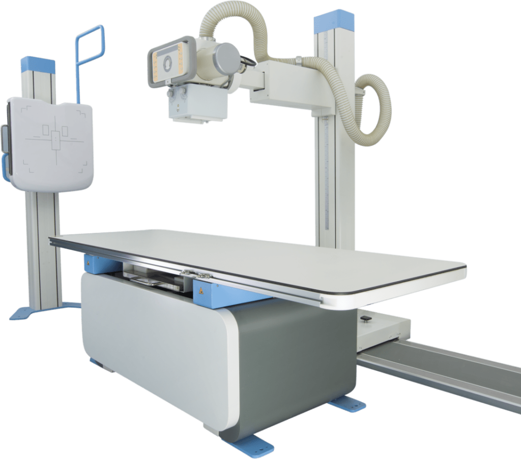
During an X-ray test, the patient is positioned on an X-ray table and an X-ray beam is directed through the body. The X-ray beam is absorbed by dense tissues such as bone, but is able to pass through soft tissues such as muscle and organs. The resulting image is captured on film or a digital detector and can be viewed on a computer screen.
X-ray tests can be done on different parts of the body, such as the chest, bones, teeth, and gut. Some examples of common X-ray tests include:
Chest X-ray: used to evaluate the lungs, heart, and other structures in the chest
Skeletal X-ray: used to evaluate bones and joints for fractures, dislocations, or other abnormalities
Abdominal X-ray: used to evaluate the organs in the abdomen
Dental X-ray: used to evaluate the teeth and jaw
X-ray tests are considered safe, but they do expose the patient to a small amount of ionizing radiation. Therefore, the test should only be done when necessary and in the lowest radiation dose possible. Pregnant women and children are more sensitive to radiation and therefore, the test should be done with caution.
Call us for an appointment
933-604-1343
Feel free to contact us.
cbsingh@ramapathology.com
Reach to us via our location
View Map